
The fabrication of removable complete dentures is a sophisticated synergy between clinical expertise and technical artistry. It is a sequence where biological assessment meets mechanical engineering. While the final aesthetic outcome is often what the patient notices, the longevity and function of the prosthesis rely heavily on the foundational steps: capturing a precise master cast and the accurate articulation of the models.
In this post, we explore the critical workflow of fabricating anatomical complete dentures, focusing on the pivotal transition from the clinical final impression to the laboratory crafting of the prosthesis.
The Clinical Foundation: Capturing the Final Accurate Die Stone Model
The journey to a successful denture begins with the accurate reproduction of the oral anatomy. The transition from a preliminary impression to a definitive one is critical. While stock trays provide a general overview, they often lack the custom adaptation required for optimal retention and stability.
The gold standard involves the fabrication of a custom tray, often utilizing light-cured resin materials. This tray is designed on a preliminary diagnostic cast, providing a “tailored fit” for the patient’s unique arch morphology.
The Critical Step: The Final Impression
The dentist uses this custom tray to capture the fine details of the residual ridge and, crucially, the peripheral borders. Border molding is performed—using heavy-body polyether or silicone compounds—to capture the functional movements of the buccal, labial, and lingual tissues. This creates the all-important peripheral seal, which is the primary retention mechanism for a complete denture.
Once the borders are defined, a light-body wash impression material is used to capture the microscopic details of the mucosa. This results in the final impression, which serves as the negative mold for the master cast.
Table 1: Clinical Stages of Final Impression
| Clinical Step | Material/Tool | Objective |
| Custom Tray Fabrication | Light-cured acrylic resin | To create a tray that fits the patient’s arch with uniform space for impression material. |
| Border Molding | Heavy-body silicone/Polyether | To record the depth and width of the vestibule and establish the peripheral seal. |
| Wash Impression | Light-body silicone/Polyether | To capture fine anatomical detail of the residual ridge. |
| Master Cast Fabrication | Type IV Dental Stone | To create the definitive working model representing the patient’s anatomy. |
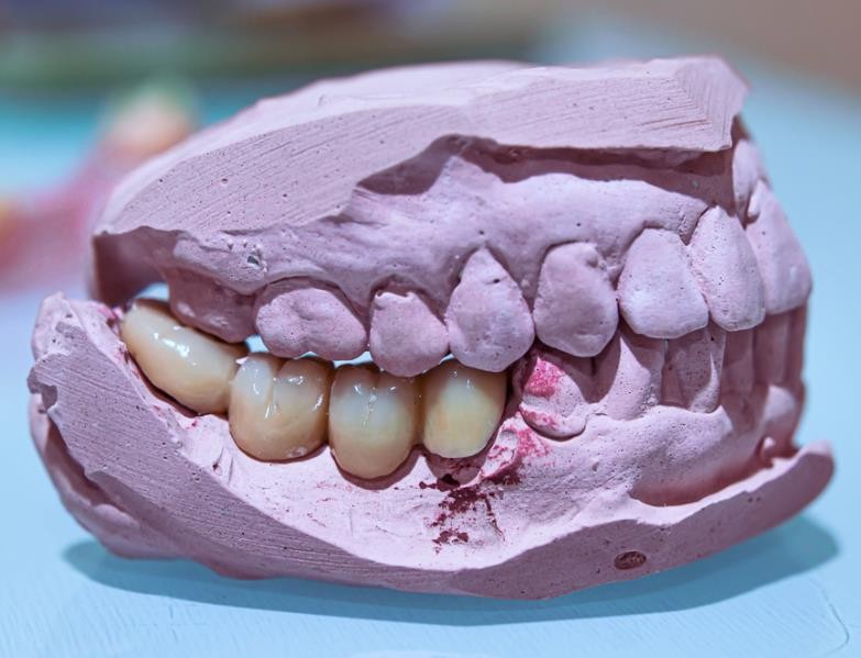
Following the impression, the Master Cast (Die Stone Model) is poured using Type IV dental stone (high-strength stone). This model must be defect-free, capturing every minute detail of the impression. It is the “ground truth” upon which the technician will build the prosthesis.
The dentist transmits the complete facebow, which incorporates the recorded articulation data and mandibular data, to the dental technician. This facilitates the accurate mounting of the maxillary and mandibular casts on the articulator, ensuring the precise reproduction of the patient’s occlusal relationship for subsequent denture fabrication.
The Laboratory Phase: Articulation and Anatomical Fabrication
Once the master cast is received in the laboratory, the technician assumes the role of an architect. The first task is to transfer the patient’s jaw relations onto an instrument that mimics the patient’s temporomandibular joint (TMJ).
Mounting on the Articulator
The technician uses a facebow record (transferred clinically) to mount the maxillary cast onto a semi-adjustable articulator. This aligns the maxilla relative to the hinge axis of the skull. Subsequently, the mandibular cast is mounted using a centric relation record. This step ensures that the artificial teeth will occlude in harmony with the patient’s jaw movements.
Arranging Anatomical Teeth
The heart of the fabrication process is the arrangement of denture teeth. Unlike flat or non-anatomical teeth, anatomical teeth mimic the natural occlusal morphology of human dentition. The technician must balance two opposing forces:
- Esthetics: Positioning teeth to support the lips and facial muscles, restoring the patient’s natural contour.
- Function: Establishing balanced occlusion—contacts that are stable during both static closure (Centric Occlusion) and dynamic movements (lateral and protrusive excursions).
The technician meticulously carves the baseplate wax to simulate the contour of natural gingiva, often referred to as “gingival festooning.” This creates a lifelike appearance, tinting the acrylic to match the patient’s natural tissue tones.
Table 2: Laboratory Fabrication Workflow
| Laboratory Stage | Procedure | Key Considerations |
| Cast Preparation | Boxing and pouring impressions; base formation. | Elimination of air bubbles; preservation of vestibular depth. |
| Articulation | Facebow transfer and mounting. | Accurate reproduction of the condylar inclination and Bennett angle. |
| Teeth Setup | Arrangement of anatomical denture teeth. | Achieving bilateral balanced occlusion; neutral zone placement. |
| Wax Try-in | Wax-up contouring and tinting. | Simulation of natural gingival texture (stippling) and root eminences. |
| Processing | Flasking, boil-out, packing, and curing (Heat-cure acrylic). | Managing polymerization shrinkage; ensuring proper curing cycle. |
The Transformation: From Wax to Resin
After the “Try-in” appointment, where the dentist verifies the esthetics and occlusion intraorally, the denture returns to the lab for processing. The wax is eliminated through the “lost wax” technique (boiling out). The void left behind is packed with heat-cured polymethyl methacrylate (PMMA) resin.
The curing cycle involves heating the flask under pressure to polymerize the resin, transforming it from a dough-like state to a rigid, durable material. After curing, the denture is deflasked, finished, and polished to a high luster, ready for delivery.
Dentsma supplies essential tools and materials for removable complete denture fabrication, from dentist’s facebow and light – cured custom trays to laboratory articulators, flasks to air pressure curing unit and polishing lathe dental lab equipments, meeting both clinical and lab needs.
Conclusion
The fabrication of a removable complete denture is a meticulous journey that demands precision at every turn. The final accurate die stone model acts as the blueprint, while the articulator serves as the simulation engine. It is only through the seamless integration of the dentist’s clinical impressions and the technician’s anatomical knowledge that a patient can be restored to full function and confidence.
Table 3: Summary of Critical Success Factors
| Factor | Impact on Final Prosthesis |
| Accurate Border Molding | Determines the retention (suction) and stability of the denture. |
| Type IV Die Stone Accuracy | Ensures the denture base fits the patient’s ridge without rocking or pressure spots. |
| Correct Articulator Mounting | Prevents occlusal interferences, protecting the residual ridges from excessive load. |
| Anatomical Teeth Setup | Restores masticatory efficiency and supports facial aesthetics. |
